Presumed Microbial Keratitis With Continuous Wear Of Silicone Hydrogels: A Case Report
Dr Padmaja Sankaridurg Program Director, Myopia Program of the Vision Cooperative Research Centre (Vision CRC), Sydney, Australia
She was awarded her B.Opt from the Elite School of Optometry, Chennai, India in 1989 and in 1999 she gained her PhD degree from the University of New South Wales, Australia. After working for a number of years at the L.V.Prasad Eye Institute as the Chief of Contact Lens Services, she took up a position at Vision Cooperative Research Centre (Vision CRC). Her research areas include myopia, contact lens induced infection and inflammation of the eye. She is a member of the International Society for Contact Lens Research, the Association for Research in Vision and Ophthalmology and the International Association of Contact Lens Educators.
Pravin Krishna LV Prasad Eye Institute, Hyderabad, India
Pravin Krishna completed his basic medical education at Bangalore Medical College, and his internship at Victoria Hospitals, Bowring & Lady Curzon Hospital, Bangalore. After an MS in ophthalmology from Guntur Medical College in Andhra Pradesh, he came to LVPEI for a fellowship in Cornea and Anterior Segment and, subsequently, joined the Institute as a consultant in the cornea services. Pravin Krishna received the SERI – ARVO Young Investigator Award for outstanding research in clinical ophthalmology for 2005 . He was shortlisted for the Col Rangachary award at the All India Ophthalmological Society annual meeting in 2006 for his work on confocal microscopy. Currently, he is in charge of the Refractive Services and Contact Lens Services at LVPEI. His areas of interest include corneal surgery, cataract and refractive surgery, contact lens and corneal diagnostics. He has published several papers, and presented posters at international and national forums.
Prashant Garg LV Prasad Eye Institute, Hyderabad, India
Prashant Garg MD, is a Consultant Ophthalmologist and Director of Education at
L V Prasad Eye Institute, Hyderabad. He is also the Medical Director of Ramayamma International Eye Bank, Hyderabad. His major area of research has been corneal infection especially understanding pathophysiology and management of fungal keratitis. He is also involved in clinical trails of various drugs. Dr. Garg is active in various national and international societies. He has authored nearly 50 papers in peer reviewed journals and chapters in textbooks. He is also reviewer for various journals including Ophthalmology and British Journal of Ophthalmology. He has several research grants to his credit.
Many events of Microbial Keratitis (MK) with silicone hydrogels have now been reported in the literature.1-3 The increased oxygen transmissibility of silicone hydrogels have eliminated all problems associated with hypoxia, however the risk of serious complications such as Microbial Keratitis while wearing these lenses is still being determined.
We present a case report of an event of Microbial Keratitis that was observed with silicone hydrogels. The event was culture-negative on microscopic evaluation of corneal scrapings but had clinical features suggestive of microbial keratitis.
The patient, a 20-year-old male participated, in a clinical trial involving wear of NIGHT & DAY™ in one eye and an experimental silicone hydrogel lens on the contralateral eye. He was new to contact lens wear and had not worn any contact lenses prior to his participation in the clinical trial. The lenses were worn on a 30 night continuous wear schedule and lens power was -4.75D in the right eye and -4.50D in the left eye. Duration of lens wear in the current trial was 84 days and his current pair of lenses was 19 nights old.
The patient reported with symptoms of pain, redness and watering present in the NIGHT & DAY™ lens wearing eye since the previous night. In spite of symptoms, the patient slept with the contact lenses and noticed mild lid edema, severe redness, pain and watering on waking. He then removed and disinfected his lenses and presented to the clinic that afternoon as the symptoms worsened.
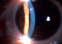
Presenting visual acuities were 6/6-3 with spectacles in both eyes and comparable to vision at baseline. Slit lamp examination revealed mild lid edema and marked injection of the bulbar and limbal conjunctiva. In the superio- nasal quadrant of the cornea, 11 0’clock meridian, there were 2 dense, yellowish -white focal infiltrates (0.5 x 0.5mm and 0.3 x 0.3mm) (Figure 1).
Figure 1
Content on this page requires a newer version of Macromedia Flash Player. Do you want to download it now?
The infiltrates were anterior stromal in depth and the larger of the two infiltrates had a full thickness epithelial defect overlying the infiltrate. There was an epithelial tag at the edge of the defect that became dislodged during examination. In addition, the infiltrates were surrounded by diffuse cellular reaction and there was mild anterior chamber reaction. Corneal scraping was performed using sterile no. 15 surgical blade on Bard Parker knife handle. The microscopic examination of smears using Grams stain and 10% KOH mount did not reveal any microorganisms. Lens wear was discontinued and treatment commenced with Ciprofloxacin 0.3% eye drops on a half hourly basis and Homide (homatropine 0.5%) eye drops thrice daily.
The following day, the patient was symptomatically better, the conjunctival redness reduced in severity and the anterior chamber reaction resolved. The clinical appearance of the corneal infiltrates, however, remained the same. Ciprofloxacin 0.3% eye drops was tapered to 2 hourly basis. On Day 2, there was further improvement in the redness levels and the density of the infiltration had also decreased (Figure 2).
Figure 2
click to enlarge
Three days later, the infiltrate began to scar at the edges. Cultures did not reveal any growth at the end of three days of inoculation. The frequency of Ciprofloxacin was reduced and the patient continued on the cycloplegic drops. Five days later, there was complete resolution of the infiltrates with scar formation. Ten days later, the patient was reviewed again and was asymptomatic and there were 2 scars corresponding to the infiltrates, measuring 0.6 x 0.5mm and 0.2 x 0.2mm (Figure 3).
Figure 3
click to enlarge
All medications were discontinued at this visit. At this visit, the patient was permanently discontinued from the clinical trial.
Discussion
Events such as Contact Lens Peripheral Ulcer (CLPU) are associated with a single, circular, circumscribed, sub-epithelial infiltrate in the periphery /mid-periphery of the cornea and appear similar to the infiltrate seen in the current event. However, the nature of the signs associated with the current event, i.e. severe bulbar and limbal redness, lid oedema, stromal infiltrate, presence of a satellite lesion and anterior chamber reaction are common features of microbial keratitis and rare in CLPUs.
The culture negative results of corneal scrapings do not rule out a microbial event. It is well established that the rates of positive cultures in cases of presumed infectious keratitis are variable 4,5 and dependent on many factors such as single versus multiple scrapes, size of the lesion, depth of lesion and media used for inoculation. Given the variability of the results of microbiology work-up, the rapid progression of the event with serious consequences and bacteria being most common organisms isolated from cases of confirmed Microbial Keratitis in contact lens wearers it is widely accepted as standard practice to initiate empirical therapy with a broad spectrum antibiotic in a situation where the diagnosis is presumed to be microbial keratitis.
Yet another feature of this event was that the patient did not present at the onset of symptoms and came in to the clinic only when symptoms worsened. In spite of repeated and careful patient instructions, one finds it a common feature in clinical practice where patients present to the clinic only upon the condition producing worsening symptoms or is no longer tolerable. Fortunately in the present case, the infiltrates were located in the mid-peripheral quadrant and did not impact vision. Practitioners should ensure that patients are instructed to return/contact their eye care professional soon after onset of symptoms if any and not delay seeking advice. When they present seeking attention for their symptoms they should be attended to promptly and appropriate action taken to: a) limit the course of the disease/condition and b) achieve the best outcome possible.
The patient was permanently discontinued from lens wear on resolution of the condition. If a patient is interested in resuming lens wear after an event, the practitioner will need to discuss the risks of recurring problems such as repeat infections prior to refitting them with contact lenses.
Whiting MAN, Raynor MK, Morgan PB et al. Continous wear silicone hydrogel contact lenses and Microbial Keratitis. Eye 2004;18: 935-937.
Schornack MM, Peterson D: Staphylococcus aureus ulcer associated with continous wear of silicone hydrogel contact lenses. Eye & Contact Lens 32
(2): 72-74, 2006.
Wong T, Ormonde S, Gamble G et al. Severe infective keratitis leading to hospital admission in New Zealand. Br J Ophthalmol. 87, 1103-1108, 2003
Lam DSC, Houang E, Fan DSP etal. Incidence and risk factors for Microbial Keratitis in Hong Kong: Comparison with Europe and North America. Eye. 2002, 16(5), 608-618.
Tell
a friend
All rights reserved, copyright 2002 - 2007 siliconehydrogels.org


 Pravin Krishna LV Prasad Eye Institute, Hyderabad, India
 Prashant Garg LV Prasad Eye Institute, Hyderabad, India