This web site is no longer actively maintained. Please visit http://www.contactlensupdate.com for up to date information.
Search
Powered by Google
Home
This Month
Editorial
Ocular Surface Characteristics of the Asian Eye
>
more
Meeting Synopsis
Academy 2010
>
more
Posters
pective Analysis of Risk Factors Associated With Contact Lens Induced Inflammatory Events During Continuous Wear
>
more
Feature Review
Adequate tear mixing under a soft contact lens may play an important role in minimizing certain > more
Tell a friend
> Home
> About Us
> Affiliates
> Contact Us
> Disclaimer
> Site Map


The Silicone Hydrogels website is partially supported through an educational grant from CIBA VISION

 
Editorial | Previous Editorials
April 2002


CLINICAL PERFORMANCE AND OBSERVATION: CORNEAL EDEMA

Deborah Sweeney -
BOptom (UNSW) 1980 PhD (UNSW) 1992

Deborah Sweeney is Professor and Chief Executive Officer of the Vision Cooperative Research Centre.  Her major research area has been corneal physiology, and her work has been instrumental in developing an understanding of the physiology of the human cornea and the effects of contact lens wear on corneal function characteristics.  Associate Professor Sweeney is also active in national and international optometric and ophthalmic organizations, including Executive roles in the International Society for Contact Lens Research, The Keratoprosthesis (KPro) Study Group and the International Association of Contact Lens Educators.


Introduction
Overnight Edema Levels
Striae and Folds
Microcysts
Limbal Redness Reduction and Ghosting of Vessels
Summary
References


Introduction
High Dk soft contact lenses (SCLs) have now been on the market in some countries for over two years and researchers have been appraising the effects of these new and innovative procedures for many more years.

From all this experience it is very clear that the hypothesis that, if materials met the Holy Grail and had a Dk/t of 87 barrers (1) or more, the materials would eliminate the hypoxic stress associated with conventional lenses for extended wear, has been proved. 

A number of clinical signs may be observed with silicone hydrogels lenses, and it is useful for practitioners to recognise these signs and their causes.    

Overnight Edema Levels

As Kathy Dumbleton reviewed for us in January, studies by CCLR clearly demonstrated that central corneal swelling was significantly reduced with these materials compared to what is found with conventional hydrogels used for extended wear ("The Physical and Clinical Characteristics of Silicone Hydrogel Lenses: How They Work")  Overnight edema levels with silicone hydrogels are in the same order as what has been observed with no lens wear. 

Striae and Folds
With this reduced overnight corneal swelling response and the rapid de-swelling that follows on eye opening, as practitioners, we do not expect to observe either striae or certainly folds in any of our patients wearing these lenses on an extended or continuous wear basis, even if we observe them immediately upon eye opening. 

A

B

Figure 1. Stromal Striae (A) and Folds (B) (courtesy Vision Cooperative Research Centre (VisionCRC) Grading Scales)


However, with the increased power range in which both products are now available, we still need to be vigilant.

Table 1: Parameters available for silicone hydrogel materials

Proprietary Name

<PureVision

<Focus® NIGHT & DAYT

Manufacturer

<Bausch & Lomb

,CIBA Vision

Ct (@ -3.00D) mm

<0.09

<0.08

Diameter mm

<14

<13.8

Base Curve(s) mm

<8.6

<8.4, 8.6

Power D

<+6.00 to -9.00

<-0.50 to -6.00 (0.25 steps)

<+0.25 to +6.00 (0.25 steps)

<-6.50 to -12.00 (0.50 steps)

<+6.00 to -10.00

<-0.25 to -8.50 (0.25 steps)

<+0.25 to +6.00 (0.25 steps)

<-8.50 to -10.00 (0.50 steps)


Grant and Long have looked at the oxygen transmissibility profile of siloxane hydrogel contact lenses and have demonstrated the effect of considering the total lens thickness profile across the whole lens diameter rather than just the central thickness of lenses ("The oxygen transmissibility profile of siloxane hydrogel contact lenses") (2).  With higher minus lenses now available and the expansion of the power ranges to also cover hyperopic corrections, it is possible that we will observe striae in some of our patients if observed soon after eye opening. A large inter-subject variability found in corneal swelling with conventional lenses has also been reported with silicone hydrogels (3). La Hood and Grant (4) have provided practitioners with an excellent method of being able to approximate the level of edema a patient is experiencing by counting the number of striae and folds. Their study showed that one striae was equivalent to approximately 5% mean corneal swelling whilst five striae equated to 11% swelling, and one fold was equivalent to approximately 8% swelling.  

There have also been a number of very interesting recent results in studies about the effect of these high Dk SCL materials on epithelial thickness as well as on stromal keratocyte density. Perez-Gomez (5) and colleagues have reported a similar reduction in posterior keratocyte density with both Acuvue 2 and PureVisionT across a six month wear period in neophytes, despite significantly less corneal swelling with the PureVisionT lenses.  This suggests that edema is not driving all the morphological changes that accompany lens wear of these novel materials and that some effects may be related to the physical presence of contact lenses and possibly a mechanical effect of contact lens wear. As suggested in the review of this month's scientific article, further investigation of these effects is needed to truly understand the mechanisms that are responsible for such changes. 

Microcysts
Another marker, perhaps the most clinically 
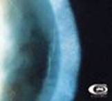
useful, is the observation of epithelial microcysts. Microcysts are small, irregularly shaped high refractive inclusions that form in the basal layers of the epithelium and move towards the anterior surface of the cornea.
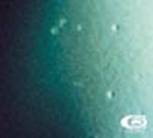
Figure 2. Corneal Microcysts (courtesy Vision Cooperative Research Centre (VisionCRC) Grading Scales)


The technique for observing epithelial microcysts uses retro-illumination. The cornea is scanned with a 1mm wide slit beam on a slit-lamp biomicroscope. A magnification of at least 16x is required with marginal retro-illumination.  Once observed, magnification is increased to 20-40x while keeping the inclusion centred in the beam and confirming that it shows reversed illumination as demonstrated in Figure 3. Microcysts are typically 10-50 microns in diameter and appear similar to micropunctate corneal staining.

Figure 3. Observation of Unreversed and Reversed Illumination (courtesy IACLE)


Relatively low numbers of microcysts are observed in patients wearing silicone hydrogels if they are new to contact lens wear or have been wearing these lenses for some time (several months or longer). This is because, as has been demonstrated previously (6), the oxygen transmissibility of lenses is inversely proportional to the number of microcysts seen.

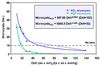
Figure 4. Relationship between microcyts and Dk/t for RGP and soft lenses (adapted from "Silicone Hydrogels: the rebirth of continuous wear contact wear" ed. Sweeney, DF)

The number of microcysts is generally less than 10, a number commonly seen in non-lens wearers and with daily wear of lenses. Large number of microcysts however will be found in a significant proportion of our patients that we transfer from low Dk lenses particularly if worn on an extended wear basis, to high Dk SCLs. In these patients, as has been reported by Keay and colleagues (7), a spike or rebound effect will be observed with large numbers of microcysts observed in the cornea for up to one month after refitting. These large numbers of microcysts are seen to be spread across the cornea or they have been noticed to aggregate in a ring in the corneal mid-periphery. As microcysts move to the anterior surface of the cornea, areas of negative staining, or black spots, may be observed (best observed after instillation of fluorescein using a cobalt blue illumination with a Wratten yellow filter).

Figure 5.  The typical trend in microcyst numbers when moving patients from low Dk to high Dk materials (adapted from "Silicone Hydrogels: the rebirth of continuous wear contact wear", ed. Sweeney, DF)

Such a trend has also been observed when patients are discontinued from low Dk extended wear. It has been suggested that the rebound is related to re-oxygenation of the corneal surface resulting in recovery of epithelial metabolism and so clearance of extra-cellular debris trapped in the deeper basal layers of the epithelium.

We have also had the opportunity in our long-term studies to monitor the levels of microcysts in our high Dk patients over a number of years and with patients on both a 6 and 30 night continuous wear schedule. We have observed no shift or increase in the low numbers of microcysts observed across time and nor have we seen any differences in the levels of microcysts observed with either a 6 or 30 night wear schedule.  

When observing the corneal epithelium it is important also to ensure that microcysts are differentially diagnosed from the many other presentations with which they may be confused. The table below adapted from Keay et al (7) details a number of conditions that can have a similar appearance to microcysts and the main distinguishing feature by which to differentiate them from microcysts.

Table 2: Differential diagnosis with other epithelial events


  <Size (µm)

Appearance

Cause

Recurrent corneal erosion

<15 to 100

Clear cysts

Trauma often unknown

Microcystic edema

<20 to 50

Clear cyst surrounding epithelial haze, > 200

Inflammatory origin

Epithelial infiltrates

<100 to 500

Granular, dense centre

Chemotactic stimulus

Vacuoles

<20 to 50

Round, bubble-like, unreversed illumination

Hypoxia

Mucin balls*

<< 100

Spherical balls

Surface interaction between lens and cornea

Punctate corneal staining

<10 to 50

Fine opaque dots, positive stain

Epithelial trauma (e.g. toxic or dehydration)

Microcysts

<10 to 50

Small, irregular-shaped dots; reversed illumination (negative stain)

Hypoxia

* embedded in epithelial surface
Limbal Redness Reduction and Ghosting of Vessels
As well as a reduction on the overall level of limbal redness that accompanies wear of silicone hydrogels, a reduction in the limbal vascularisation has also been observed. As the level of limbal redness have been associated by Papas and others (8,9) with the level of edge oxygen transmissibility, it has also been proposed that the unfilling (or ghosting) of limbal vessels is also associated with the increased oxygen transmissibility of these lenses in the peripheral area of the lens. More about the mechanism involved in reduced limbal redness as well as new clinical techniques which are being developed to monitor limbal redness will be discussed in the May editorial.

Summary
As always, as practitioners, we need to closely monitor our patients to ensure their ongoing ocular health. Silicone hydrogels bring with them their own set of observations and events as the eye adjusts to the new lenses. Being able to recognise and differentiate normal changes from adverse events or reactions with silicone hydrogels will enhance patient management and is an important part of continuous wear practice.


References

1. Holden B and Mertz G. Critical oxygen levels to avoid corneal edema for daily  and extended wear contact lenses. Invest Ophthalmol Vis Sci 1984; 25:1161-1167.

2. Grant R and Long B. The oxygen transmissibility profile of siloxane hydrogel contact lenses.  Optom Vis Sci 2000; 77(suppl):264.

3. Mueller N, Caroline P, Smyhte J, Mai-Le K and Bergemske G.  A comparison of overnight swelling response with two high Dk silicone hydrogel contact lenses. Optom Vis Sci 2001; 78(suppl):199.

4. La Hood D and Grant T. Striae and folds as indicators of corneal edema. Optom Vis Sci 1990:67(suppl):196.

5. Perez-Gomez I, Morgan PB and Efron N. Confocal microscopic appearance and thickness of the cornea following extended wear contact lenses: A study with neophytes. Optom Vis Sci 2001;78(suppl):199.

6. Holden BA, La Hood D and Sweeney DF. Prediction of extended wear microcyst response on the basis of the mean overnight corneal response in an unrelated sample of non-wearers. Am J Optom Physiol Opt 1987;64:83.

7. Keay L, Jalbert I, Sweeney DF and Holden BA. Microcysts: clinical significance and differential diagnosis. Optometry 2001;72(7):452-460.

8. Papas E, Vajdic CM, Austen R and Holden BA. High Dk soft contact lenses reduce the limbal vascular response. Optom Vis Sci 1994;71:14.

9. Dumbelton K, Richter D, Simpson T and Fonn D. A comparison of the vascular response to extended wear of conventional lower Dk and experimental high Dk hydrogel contact lenses. Optom Vis Sci 1998;75(suppl):170.

 
All rights reserved, copyright 2002 - 2007 siliconehydrogels.org