| INTRODUCTION |
| The eye has a range of natural defence
systems which very effectively protect the eye's tissues from
inflammation and infection. The placement of a contact lens on
the eye, however, places an added burden on these systems, which
can sometimes result in an adverse response or event.
In practice, being able to recognise and effectively manage adverse
events is an important part of our professional responsibilities.
Accurate diagnosis enables us to identify possible causes and
risks, to decide whether treatment is necessary and gauge the
time to resolution, and to inform the patient of the likely time
out of lens wear, chance of recurrence, and strategies to reduce
risk. In this editorial we will look at typical adverse events
that accompany lens wear, what events are occurring with silicone
hydrogel lenses, and how these events can be effectively managed.
 |
| INCIDENCE |
| The high oxygen permeability and other
physical features of the new materials, have impacted the incidence
of adverse responses seen with silicone hydrogel lenses.
The new highly oxygen permeable silicone hydrogel materials have
eliminated physiological changes due to hypoxia. Overnight edema
levels are similar to the levels seen with no lens wear and are
far lower than those with commercially available disposable soft
lenses [1][2]. Microcysts,
the main clinical indicator of corneal edema [2]
are rarely if ever seen with silicone hydrogel EW (except when
initially transferring a low Dk wearer, the "rebound effect",
this is the same phenomenon that occurs when some low Dk wearers
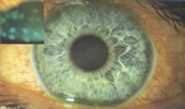
discontinue lens wear.). We also find remarkably 'white eyes'
with silicone hydrogel lens wearers due to the reduction in limbal
hyperaemia. 'Ghosting' of neovascularisation in previous wearers
of Low Dk EW soft lenses due to emptying of the vessels occurs
in silicone hydrogel lens wear [3][4].
It is hypothesised that the increased oxygen permeability of
the silicone hydrogel lenses ensures that the eye's defences are
not compromised due to a healthier epithelium, providing better
protection against infection (Microbial Keratitis).
Studies at Vision Cooperative Research Centre (VisionCRC) and CCLR of adverse responses with the
new lenses compared to low Dk lenses have shown that there are
similar rates of inflammatory conditions but higher rates of some
of the mechanically induced adverse events. |
| EVENTS |
| While clear identification of an adverse
event is essential, unfortunately widespread confusion still exists
in the literature and in clinical practice, with either the same
adverse reactions being categorised differently or different reactions
being labelled as the same type of event. For example, the term
'corneal infiltrate' is used to describe anything from a large
infected ulcer to a few cells in the cornea.
Microbial Keratitis (MK) is the only infection related to contact
lens wear. All other events are either inflammatory in nature
or mechanically induced. The inflammatory events such as CLPU,
CLARE, IK, AIK
and AI may be associated with the presence of
bacteria and their toxins on the eye, the lids or the lens, but
are not infective. Infiltrates can occur in response to infection,
inflammation and mechanical disturbance to the cornea.
Vision Cooperative Research Centre (VisionCRC) and the LV Prasad Eye Institute (LVPEI) in India have conducted
numerous clinical contact lens trials in the last 10 years. Detailed
information on the signs, symptoms, appearance, laboratory findings,
treatment and management of a range of adverse events have been
collected. This information resulted in the development of the
Vision Cooperative Research Centre (VisionCRC)/LVPEI Guide to Corneal
Infiltrative Conditions seen in Contact Lens Practice (PDF-2.24MG).
This Guide categorises events into serious and symptomatic; clinically
significant and commonly symptomatic; and clinically non-significant
and asymptomatic.
The Vision Cooperative Research Centre (VisionCRC)/LVPEI Guide to Corneal Infiltrative Conditions seen
in Contact Lens Practice can now be viewed under the new Resource
Section of the Research Library. Copies of the Vision Cooperative Research Centre (VisionCRC)/LVPEI Guide
to Corneal Infiltrative Conditions seen in Contact Lens Practice
can be obtained by contacting Vision Cooperative Research Centre (VisionCRC) (k.evans@visioncrc.org)
Below is some information about particular adverse events: |
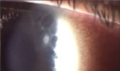
| MICROBIAL KERATITIS
(MK) |
| - serious and symptomatic
Microbial Keratitis is caused by microbial infection of the cornea,
and is classified as a serious adverse event. It is has the potential
to result in vision loss, although the reports of cases resulting
in vision loss are extremely low.
MK is rare and is the only serious adverse event to occur
in contact lens wear.
The presentation of MK can vary depending on the type and virulence
of the micro-organism involved, and the stage at which the patient
presents. In general, excavation of the corneal epithelium, Bowman's
layer and stroma is seen, with serious necrosis and infiltration
of the underlying tissue [5]. The shape of
the lesion is usually irregular and "satellite" lesions
(smaller lesions adjacent to the primary site of infection) may
be present. Anterior chamber reaction is often observed in the
active stage. |
| |
| Aetiology: Risk factors
such as hypoxia, trauma, ocular surface disease, certain systemic
conditions and contact lens wear are known to be associated with
the development of MK [6][7][8][9][10].
Treatment: Prompt attention and treatment are mandatory
as infection associated with virulent organisms can progress and
cause severe destruction of the cornea within 24 hours. Conjunctival
swabs and a corneal scrape will help to determine the type of
microbe involved. Aggressive antibiotic therapy delivered topically
at frequent intervals is then the usual approach, however treatment
can vary according to the stage and severity of the condition.
Once the microbe has been identified the treatment may be modified
based on the sensitivity of the organisms to particular antibiotics.
While MK is rare, significant delay or inappropriate treatment
can affect the visual outcome. A conservative approach is therefore
recommended with the treatment of any focal infiltrate with significant
overlying staining, until the possibility of MK has been eliminated. |
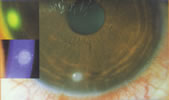
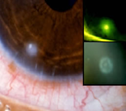
| CONTACT LENS
INDUCED PERIPHERAL ULCER (CLPU) |
- clinically significant and commonly
symptomatic
A circular, well circumscribed, dense, yellowish-white, focal corneal
infiltrate (0.2-2.0mm in diameter) located in the peripheral to
mid-peripheral cornea, with diffuse infiltration of the surrounding
stroma. In its active stage, CLPU is characterised by marked limbal
and bulbar redness, usually localised to the quadrant adjacent to
the lesion. This is the most common symptom reported by the patient,
followed by pain or soreness, irritation or watering. CLPU is usually
unilateral and typically features a single focal infiltrate, however
a number of events can occur, although this is rare. In severe cases,
there may be mild anterior chamber involvement and photophobia.
|
| |
| Aetiology: CLPU is an
inflammatory reaction of the cornea thought to be due to bacteria
adhering to the contact lens or toxins released by the bacteria.
The cornea is not infected and bacteria is not found on scraping
or biopsy of the lesion.
Treatment: In its acute phase CLPU necessitates
temporary discontinuation of lens wear until resolution of the
corneal infiltrates. No medical therapy is needed. Patients should
be monitored until the event resolves. The events always resolve
in a characteristic 'bulls-eye' scar. The patients that present
to our clinics with healed peripheral circular scars without treatment
highlight the self-limiting nature of CLPU events. The condition
is prone to recurrence but does not necessitate permanent discontinuation.
CLPU can mimic early MK, however with CLPU the symptoms
are milder and begin to recede immediately on discontinuation
of lens wear. Again, a conservative approach is recommended however
with the treatment of any focal infiltrate with significant overlying
staining, until the possibility of MK has been eliminated. |
| CLPU
vs MK |
 |
 |
| CLPU:
On removal of lenses there is rapid, uncomplicated resolution
resulting in a small “bullseye” scar |
MK:
Worsens without aggressive treatment, stromal tissue destruction
and scarring |
|
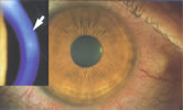
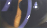
| CONTACT LENS INDUCED
ACUTE RED EYE (CLARE) |
- clinically significant and commonly
symptomatic
CLARE is an inflammatory reaction following overnight sleep with
contact lenses. It only occurs after sleep and is usually unilateral.
The patient typically presents with marked conjunctival redness
and irritation or pain. Approximately one third of CLARE patients
in our experience are woken by severe pain. Other symptoms noticed
on waking or shortly after included discomfort and watering. The
most characteristic feature of the condition is the presence of
fine diffuse cellular infiltration of the peripheral to mid-peripheral
cornea with clusters of small focal infiltrates interspersed or
extending into the clear cornea. The conjunctival redness and infiltrates
is circumferential in nature, the infiltrates appearing to "stream"
from the limbal vessels. If infiltrates extend into the pupillary
zone, photophobia may be experienced by the patient. The extent
of involvement ranges from 10-360 degrees of the corneal circumference,
although most commonly three quarters are affected. There is sometimes
epithelial involvement, but if present it is limited to minimal
punctate corneal staining over the infiltrates. The infiltrates
are found in the anterior stroma, and the posterior stroma and anterior
chamber are unaffected. |
| |
| Aetiology: A significant
number of lenses recovered from cases of CLARE have been contaminated
with Gram negative bacteria. It is thought the reaction is an
inflammatory response to the toxins produced by the bacteria.
Treatment: Temporary discontinuation of lens wear
until resolution of the corneal infiltrates is required. This
may take one to two weeks. As with CLPU, CLARE simply resolves
on cessation of lens wear without the need for medical therapy.
Patients should be monitored until the event resolves. Approximately
one third of the cases recur [11][12],
and patients need to be counselled as to the possibility of recurrence
before recommencing lens wear. |
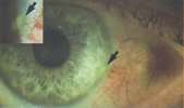
| INFILTRATIVE KERATITIS
(IK) |
- clinically significant and commonly
symptomatic
IK is a general category for symptomatic infiltrative events in
contact lens wear that do not fit the profile of MK, CLPU or CLARE.
Typically IK is a relatively mild event characterised by anterior
stroma infiltration with or without epithelial involvement in the
periphery to mid-periphery of the cornea. IK is usually accompanied
by moderate redness and irritation. As with CLPU and CLARE, photophobia
may be a symptom if the infiltrates extend beyond the pupil margin.
CLPU can easily be distinguished from IK by its very distinctive
round lesion. CLARE occurs only after overnight wear and patients
are generally more symptomatic. |
| |
| Aetiology: IK is multifactorial
in nature. It may be in response to bacterial toxins, or a trapped
foreign body or due to mechanical trauma to the cornea. Mechanical
trauma is diagnosed by a positive history and or/an epithelial
defect suggestive of trauma.
Treatment: Lens wear should be suspended, and patients
monitored until the event resolves. Prophylactic antibiotics can
be prescribed if there is any loss of epithelium. |
| CONTACT LENS PAPILLARY
CONJUNCTIVITIS (CLPC) |
- clinically significant and commonly
symptomatic
CLPC is commonly referred to in the literature as Giant Papillary
Conjunctivitis (GPC). Studies at Vision Cooperative Research Centre (VisionCRC) indicates that there may
be two distinct categories of CLPC - general and localised. As these
two categories are thought to have different aetiologies, patient
symptoms and treatment strategies, they will be discussed separately. |
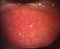
| GENERAL
CONTACT LENS PAPILLARY CONJUNCTIVITIS
(CLPC) |
| General CLPC is the form with which most
practitioners are familiar. It involves large, raised papillae of
a cobblestone appearance with moderate to severe hyperaemia across
the entire tarsus. General CLPC is characterised by moderate to
severe patient symptoms, including itching or irritation, a stringy
or ropy discharge, excessive movement of the lens and blurred vision
due to this movement or coatings/discharge on lenses. Similar levels
of generalised CLPC are found in silicone hydrogel CW and low Dk
EW. The time to occur in silicone hydrogel lens wear is on average
11 months ranging from 6-17 months [13]. |
| |
| Aetiology: The aeitiology
is uncertain, but it has been hypothesised that it is a hypersensitivity
reaction, either delayed [14] or immediate
[15][16]. It is believed
that lens deposits such as protein is the major risk factor [17][18][19].
Lower patient age [20], increased periods
of lens wear [18], infrequent replacement
of lenses [18] and the wear of larger lenses
[18] have also been suggested as risk factors.
Mechanical irritation due to front surface deposition may also
play a role.
Treatment: Management options for General CLPC
include frequent cleaning and replacement of lenses to reduce
deposits, a decrease in wear time, or a change in mode of wear
(EW to DW), lens type or material. Mast cell inhibitors in conjunction
with steroids are used in some cases to manage recurrent events.
Tarsal redness decreases significantly, but does not tend to return
to baseline levels, despite successful management. Papillae also
may remain dispersed over the entire tarsus, but are significantly
smaller. |
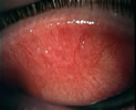
| LOCALISED
CONTACT LENS PAPILLARY CONJUNCTIVITIS (CLPC) |
| Localised CLPC involves papillae and hyperaemia
confined to one or two areas of the upper tarsus only, usually in
the central region nearest the lid margin [13].
The symptoms in Localised CLPC can be much milder than in General
CLPC, with slight irritation or foreign body sensation often the
only symptom. Preliminary evidence indicates that patients wearing
silicone hydrogel lenses are more disposed to Localised CLPC than
hydrogel lens wearers. The time to occur varies considerably with
presentation sometimes happenening very quickly after commencing
silicone hydrogel lens wear, but in other cases no significant papillae
are observed for at least two years of lens wear. |
| |
| Aetiology: Local enlarged
papillae are also found in patients with sutures, ocular prostheses
etc, suggesting that mechanical trauma has a role in the aetiology
of local CLPC [13].
Treatment: Following discontinuation of lens wear,
the tarsal redness usually resolves in 2-4weeks, however the papillae,
while smaller, tend to remain. Localised CLPC has a tendency to
recur in approximately 50% of cases [13]
and following a second episode in the one eye, current management
strategy is to change to frequent replacement low Dk daily wear
or daily disposable lens wear, which is usually successful. Refitting
with a steeper base curve may be beneficial for some patients. |
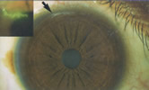
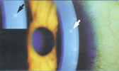
| SUPERIOR EPITHELIAL
ARCUATE LESION (SEAL) |
- clinically significant and commonly
symptomatic
SEALs present as a thin white arcuate lesion in the superior cornea,
with significant overlying staining and possible underlying diffuse
infiltrates. The edges of the lesion are often irregular and may
be slightly roughened or thickened. Approximately one third are
asymptomatic in silicone hydrogel EW, with the most common symptom
when present being foreign body sensation or irritation. Approximately
40% have underlying infiltrates and a third exhibit stromal glow
of fluoroscein. In silicone hydrogel EW, SEALs tend to occur in
two locations: limbal (immediately adjacent to the limbus) and paralimbal
(approximately 1mm from the limbus). The paralimbal lesions are
more likely to be associated with infiltrates and cause symptoms
[21]. Time of occurrence of the first event
varies widely, for example in studies at the Vision Cooperative Research Centre (VisionCRC), the time of
onset has ranged from 1-20 months. |
| |
| Aetiology: SEALs have
a multifactorial aetiology. Identified risk factors include: steep
corneas, tight eyelids, Asian eye shape, presbyopia and male gender.
Current findings of poor wettability and tight fitting lenses
in silicone hydrogel EW SEALs cases, support the hypothesis that
mechanical chaffing due to a thinning tear film in the superior
cornea are factors in the development of SEALs.
Treatment: Discontinuation of lens wear until resolution
of the SEAL, including infiltrates, is recommended. Resolution
usually occurs within 24-48 hours, but may take 1-2 days longer
if infiltrates are present. SEALs tend to recur in approximately
50% of cases, but the time to recur varies widely among patients.
There is usually no scarring on resolution or after repeated events.
|
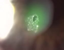
| CORNEAL EROSION |
- clinically significant and commonly
symptomatic
Corneal erosions or abrasions can occur due to mechanical trauma
(fit, lens defect, trapped foreign body or on insertion and removal)
or as a result of physiological damage due to over-wear. The damage
is usually limited to anterior to Bowman's layer. The signs and
symptoms can vary widely depending upon the cause. If the depth
of the erosion is limited to the superficial 1-3 layers of the epithelium,
the event is often asymptomatic however, if it is deeper, moderate
to severe pain, watering and blepharospasm may be present [22].
If infiltrates are present, the event becomes infiltrative keratitis,
is categorised as such with the appropriate management strategy
followed. |
| |
| Treatment: If the erosion
is small (<0.5mm) and superficial, lens wear should be discontinued
for 12-24 hours and the patient should be monitored. For large
(>0.5mm) superficial or deep erosions, lens wear should be
discontinued for at least 24 hours and not resumed until complete
resolution. It is important not to patch the eye as this significantly
increases the risk of secondary infection [23].
Prophylactic antibiotics may be used in severe cases, however
preservatives in these solutions can delay healing. Artificial
tears can be used if discomfort is present. |
| ASYMPTOMATIC INFILTRATIVE KERATITIS (AIK)
& ASYMPTOMATIC INFILTRATES (AI) |
- clinically
non-significant and asymptomatic
AIK and AI are asymptomatic, and resolve rapidly on discontinuation
of lens wear. They feature small focal infiltrates and mild diffuse
infiltration. Up to 30% of the population will exhibit generally
asymptomatic, infiltrative reactions, without contact lens wear
or any other obvious aetiological factors [24][25][26].
AIK is distinguished from AI by the presence of redness and or overlying
corneal staining. The infiltrates are also usually slightly larger
than in AI (approximately 0.4mm vs 0.2mm). |
| |
| |
| Treatment: Discontinuation
of lens wear in AIK is usually recommended until the redness and/or
staining has resolved, which usually takes a couple of days. If
there are a small number of focal or only slight diffuse infiltrates
and no staining or redness, lens wear can be safely maintained.
|
DIFFERENTIAL
DIAGNOSIS:
CONDITIONS NOT ASSOCIATED WITH CONTACT LENS WEAR |
| VIRAL KERATOCONJUNCTIVITIS
(VK) |
- clinically significant and commonly
symptomatic
Viral Keratoconjunctivitis (VK) is not caused by CL wear, but can
sometimes be confused with IK. VK is characterised by multiple focal
corneal infiltrates, and irritation, redness, tearing and photophobia
on the part of the patient. The condition may be highly infectious
but is always self-limiting. Compared to IK, there are usually larger
numbers of infiltrates, although they tend to be smaller and are
often epithelial rather than stromal, and it takes longer to resolve. |
| |
| Aetiology: VK is an
immune response of the cornea, caused by various strains of adenovirus.
Treatment: Lens wear should be discontinued for the course
of the infection, which generally lasts for several weeks. Some
VK can be very contagious and caution should be exercised with
hygiene. A topical antibiotic can prevent an opportunistic, superimposed
bacterial infection. |
| ADVERSE EVENTS IN PRACTICE |
| The adverse events seen in silicone
hydrogel practice are also seen in other modes of wear. The procedures
for silicone hydrogel patient education and management should
therefore be no different to other contact lens practice.
Patients should be educated and regularly reminded that they
should contact an eyecare practitioner immediately upon noticing
symptoms. Patients should also be taught how to avoid risks with
their contact lens wear. Most importantly, they should check every
day whether their eyes look good, feel good, and see well; and
they should never wear a lens overnight if it is uncomfortable
or if they are unwell. All contact patients should have an up
to date pair of spectacles, should an adverse event necessitate
temporary discontinuation of wear.
Practitioners should ensure that they are familiar with the signs,
symptoms and management of the various types of adverse event
seen in contact lens wear, and have systems in place to provide
immediate care to any patient who suspects an adverse event. With
practitioner and patient education, adverse events can be managed
easily and effectively leading to increased patient confidence.
|


 Deborah
Sweeney - BOptom (UNSW) 1980 PhD (UNSW)
1992
Deborah
Sweeney - BOptom (UNSW) 1980 PhD (UNSW)
1992